
What Is a Stress Fracture and How Is It Diagnosed?
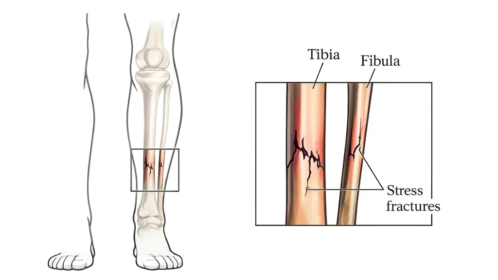
A stress fracture is a small crack in a bone that develops from repetitive force, overuse, or a sudden increase in physical activity. Doctors diagnose this stress injury through a detailed medical history, a physical exam, and imaging tests such as X-rays, magnetic resonance imaging (MRI), or bone scans, if needed.
At Total Orthopedics and Sports Medicine, our surgeon, Dr. Charles Ruotolo, uses advanced diagnostic techniques to detect early stress fractures. He reviews your symptoms, activity level, and physical exam findings before ordering imaging tests for an accurate diagnosis. This thorough approach supports early diagnosis, guides effective stress fracture treatment, and reduces the risk of severe damage. Call 855-321-6784 to get a proper diagnosis in Syosset.
How Do Doctors Diagnose a Stress Fracture?
Doctors perform a targeted physical exam and order imaging scans to diagnose a stress fracture. During the assessment, they carefully review your medical history, evaluate your symptoms, and discuss your activity level. They also assess your range of motion, walking pattern, weight-bearing ability, muscle strength, and overall limb alignment to identify factors contributing to the injury.
Moreover, doctors press on the painful area to check for swelling, bruising, and pinpoint tenderness over the affected bone. To confirm their findings and identify signs of a stress fracture, they may also perform the following physical tests:
- One-Leg Hop Test: Research published in the SLACK Journal reports that hopping on the affected leg may trigger pain that points to a stress fracture.
- Fulcrum Test: Pressure applied to the thigh may trigger pain and help identify a femoral stress fracture.
- Heel Percussion Test: Gentle tapping on the heel creates vibrations that may trigger pain at the fracture.
Which Imaging Tests Are Used to Diagnose a Stress Fracture?
Doctors first recommend X-rays to diagnose stress fractures. However, X-rays often fail to show stress fractures in the first few weeks after pain begins. In such cases, an MRI scan and computed tomography (CT scan) are used to diagnose stress fractures, especially if symptoms persist or the injury is difficult to identify. These scans provide detailed images of the bone and can detect cortical abnormalities (changes in the outer layer of the bone) that may not appear on routine imaging.
In severe cases, doctors may recommend the following diagnostic imaging tests based on the injury location, symptom severity, and the level of detail needed to confirm the diagnosis:
Bone Scan (Scintigraphy)
Doctors may recommend a bone scan to detect stress fractures that do not appear on X-rays. During the test, a small amount of radioactive tracer is injected into the bloodstream and absorbed by areas of active bone repair. These areas absorb more tracer and appear brighter on the scan, which can help identify a stress fracture. However, bone scintigraphy findings cannot always distinguish a stress fracture from other conditions, such as a high ankle sprain, infection, or tumor. Therefore, doctors interpret bone scan results alongside other imaging modalities for stress fracture diagnosis.
Ultrasound
Ultrasound uses high-frequency sound waves to examine the outer surface of the bone and nearby soft tissues. It can help identify early tibial stress injuries and detect changes around the bone before they become visible on X-rays. These changes may include tissue swelling and thickening around the injured area. However, the test’s accuracy often depends on the technician’s experience and the location of the injury.
What Are the Common Symptoms of an Overuse Bone Injury?
You may experience localized pain, tenderness over one specific spot, and mild swelling near the affected bone. You may also notice a dull ache that worsens with physical activity and improves with rest. As the stress injury progresses, the pain worsens with weight-bearing activities like walking, persists for longer periods, and interferes with normal movement. Over time, you may notice the following additional stress fracture symptoms:
- Pain at Rest or Night: Discomfort continues even when you are not active
- Joint Stiffness: Reduced movement in the nearby joint
- Muscle Tightness: Tightness around the injured area
- Limping: Changes in walking pattern to avoid pain
- Reduced Mobility: Difficulty performing daily activities comfortably
- Increasing Leg Pain: Symptoms become more noticeable during routine movement
Who Is at Risk for Developing a Stress Fracture?
Athletes and individuals with underlying bone conditions face a higher risk of developing stress fractures. Military recruits also face a higher risk due to repetitive marching, running, and load-bearing activities. In fact, research published in the British Journal of General Practice found that stress fractures occur in 5% to 20% of military personnel.
Moreover, a PubMed study found that females had higher stress fracture rates in both military and athletic populations. The researchers reported stress fracture rates of approximately 9.2% in female military personnel compared with 3% in males. Beyond that, several activities, medical, and biomechanical factors can increase the risk of these injuries, such as:
- High Exercise Volume: Repeated training places ongoing stress on the bones
- Low Calcium and Vitamin D Levels: Poor bone health increases fracture risk
- High-Impact Sports: Running, basketball, gymnastics, and similar activities increase repetitive stress
- Hard Surfaces: Training on concrete or other hard surfaces increases impact forces
- Improper Footwear: Worn or unsupportive shoes reduce shock absorption
- Osteoporosis: Reduced bone strength increases the risk of fractures
- Muscle Fatigue: Tired muscles absorb less shock and transfer more force to the bones
How Do Training Errors Contribute to Bone Stress Injuries?
Training errors alter normal body mechanics and place excessive stress on the bones. For example, sudden increases in training intensity can overload the skeleton and contribute to stress injuries. Similarly, inadequate recovery and muscle fatigue can repeatedly stress the same bone. As a result, the natural bone repair process cannot keep up with the ongoing stress. This imbalance causes microscopic bone damage to accumulate and weakens the bone over time. As these changes continue, stress reactions can progress into small bone cracks and eventually a severe fracture.
What Are the Different Types of Stress Fractures?
Tibial stress fractures, metatarsal stress fractures, and navicular stress fractures are common types of injuries that develop from repetitive stress on the bones. You may also suffer from calcaneal stress fractures (heel bone) and femoral stress fractures (thigh bone), especially during high-impact activities. Beyond this, fibular stress fractures and pelvic stress fractures can occur in people who place repeated stress on the lower body over time.
Here are some other types of stress fractures:
- Spondylolysis: A stress fracture affecting the vertebrae in the lower spine
- Fatigue Fractures: Affect healthy bones exposed to repeated stress
- Insufficiency Fractures: Affect weakened bones with reduced strength
- High-Risk Stress Fractures: These injuries occur in areas such as the anterior tibia, with poor blood supply
- Bilateral Stress Fractures: Occur on both sides of the body and account for about 16% of cases
How Often Do Stress Fractures Occur in the Shin and Pelvis?
The tibia (shinbone) is one of the most common locations for stress fractures and accounts for approximately 23.6% of cases. In contrast, pelvic stress fractures are much less common and account for about 1.6% of cases. Although pelvic injuries occur less frequently, they often require careful evaluation due to their location and potential complications, such as:
- Persistent hip or groin pain that limits walking and daily activities
- Difficulty bearing weight on the affected side
- Fracture progression into a more serious pelvic injury
- Delayed healing due to continued stress on the pelvis during movement
- Changes in walking mechanics that place stress on nearby joints and muscles
How Do Stress Fractures & Shin Splints Differ?
A stress fracture is a small crack in the bone that causes highly localized pain and often worsens with continued activity. In contrast, medial tibial stress syndrome (shin splints) causes inflammation of the muscles and connective tissues around the shinbone, often resulting in widespread pain. Moreover, shin splints may improve with warming up or rest, whereas a stress fracture may persist at night. While both conditions are overuse injuries, here are the key differences:
| Shin Splints (Medial Tibial Stress Syndrome) | Stress Fracture |
|---|---|
| General soreness along the shin | Severe pinpoint tenderness |
| Swelling is usually uncommon | Swelling may develop around the injured area |
| Often heals within 2–6 weeks | Commonly requires 6–12 weeks or longer |
| Caused by soft tissue irritation | Caused by a structural crack in the bone |
| Usually less severe | Higher risk of fracture progression |
What Treatment Options Are Recommended for Stress Fractures?
Our surgeon, Dr. Charles Ruotolo, often recommends activity modification, pain control such as acetaminophen and NSAIDs, to support stress fracture healing. He may also recommend ice therapy, compression, and elevation to reduce swelling and protect the injured bone. As healing progresses, low-impact exercises such as stationary cycling and walking boots may help maintain fitness without placing excess stress on the bone.
In severe cases, Dr. Charles Ruotolo may recommend stress fracture surgery if the fracture does not heal properly or affects high-risk areas. Here is how these treatments help promote healing, restore safe movement, and reduce the risk of future complications:
Activity Modification and Rest
Our orthopedic specialist, Dr. Charles Ruotolo, guides you to avoid running, jumping, and other high-impact activities for several weeks. He may also recommend a walking boot or crutches to reduce stress on the affected limb. This activity modification helps reduce pressure on the injured bone and supports natural healing. As a result, the bone can begin to heal properly, and the risk of the complete break decreases.
R.I.C.E. Protocol
The R.I.C.E. protocol helps control pain and swelling during the early stages of recovery. For example, rest reduces stress on the injured bone, while ice helps reduce inflammation and discomfort around the injury site. Ice also constricts local blood vessels, which limits swelling and helps reduce secondary tissue irritation around the fracture. Beyond this, compression and elevation help control fluid buildup, reduce swelling, and support comfortable recovery.
Pain Management
According to the American Academy of Orthopedic Surgeons, nonsteroidal anti-inflammatory drugs, such as ibuprofen or naproxen, may help relieve pain and reduce swelling. If pain persists in a specific area, lidocaine patches may provide temporary relief around the injury site. However, long-term use of these medications may slow stress fracture healing and delay recovery. For this reason, you should discuss pain management options with your doctor before using them regularly.
Orthotics and Supportive Footwear
Supportive footwear reduces impact on weight-bearing bones during recovery. For example, shock-absorbing insoles can help distribute pressure more evenly across the foot. Similarly, stiff-soled shoes may limit excessive movement around the injury site. As a result, the healing bone experiences less repetitive stress, which supports faster recovery after a traumatic event.
Exercises
Once the bone heals, you should perform strengthening and balance exercises to rebuild strength and support a safe return to physical activity. These exercises improve flexibility, balance, and movement control throughout the lower limbs. As strength improves, you can perform the following exercises to support long-term recovery and lower the risk of future stress injuries:
- Calf and Soleus Raises: Strengthen the lower leg and reduce stress on the feet and shins
- Glute Bridges and Hip Thrusts: Improve hip stability and reduce pressure on the lower limbs
- Squats and Lunges: Build full-leg strength and improve balance during movement
- Single-Leg Deadlifts: Correct muscle imbalances and strengthen the hamstrings and stabilizing muscles
Surgical Treatment
Our orthopedic surgeon, Dr. Charles Ruotolo, may recommend stress fracture surgery if the broken bone does not heal with conservative treatment. He may also recommend surgery for high-risk fractures with poor blood supply or a higher risk of complications. During the procedure, he may use screws, plates, or pins to stabilize the injured bone. This added stability supports stress fracture healing and lowers the risk of a complete break. Call 855-321-6784 to book an appointment for advanced treatment.
How Do Bracing and Protective Devices Support Healing?
Bracing and protective devices support healing by stabilizing injured joints and reducing stress on damaged tissues. They also provide compression, which helps control swelling and improve comfort during recovery. In addition, these devices help maintain proper alignment and protect the injured area from further strain. As a result, you can move more safely while the injured bone or soft tissues recover.
In fact, a PubMed study evaluated 42 athletes with lower-leg pain, including 17 athletes diagnosed with a tibial stress fracture. All athletes used a pneumatic leg brace on the affected limb during recovery. Researchers found that athletes with stress fractures resumed light training on average within 1 week and returned to intensive training within about 3.7 weeks. They also returned to competition at their preinjury level, on average, within 5.3 weeks.
How Long Does It Take to Recover from Stress Fractures?
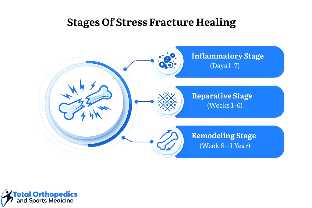
Most stress fractures heal within 6 to 8 weeks with proper rest and activity modification. However, the overall recovery time varies depending on the location and severity of the injury. For example, tibial and youth stress fractures heal within 4 to 6 weeks because these bones have a good blood supply. In contrast, high-risk traumatic fractures in areas with limited blood supply may require 3 to 6 months or longer to heal completely. The overall recovery time may also extend due to the following factors:
- Returning to Activity Too Early: Continued stress can worsen the fracture and delay healing
- Low Vitamin D Levels: Insufficient vitamin D can slow bone repair
- Poor Calcium Intake: Bone healing may take longer without adequate nutrients
- Smoking: Nicotine reduces blood flow to the healing bone
- Delayed Diagnosis: A late diagnosis of stress fractures may allow the injury to worsen
- High-Risk Fracture Location: Bones with poor blood supply generally heal more slowly
- Poor Nutrition: Low-calorie intake can interfere with bone remodeling
- Underlying Health Conditions: Certain medical conditions can delay stress fracture healing
What Signs Indicate Complete Healing?
Once the bone heals, you may notice pain-free walking and a full pain-free range of motion. As your recovery progresses, the injured area should no longer feel tender, and everyday activities such as climbing stairs should become comfortable again. You should also perform these activities without swelling or discomfort around the injured bone. However, to confirm complete bone healing, your doctor may order follow-up imaging, such as X-rays or computed tomography (CT) scans.
Can Stress Fractures Recur After Treatment?
Yes, some people experience recurrent stress fractures even after recovery. This often happens when you return to high-impact activity before the bone has fully healed. Muscle imbalances, poor movement mechanics, overtraining, or worn footwear can also place repeated stress on the injured bone. Beyond this, low vitamin D levels, inadequate calcium intake, and underlying bone weakness can further increase the risk of future stress injuries.
How Can You Prevent Future Stress Injuries?
To reduce the risk of future stress injuries, gradually increase training loads and maintain adequate vitamin D and calcium intake. You should also engage in low-impact cross-training and wear supportive footwear to reduce repetitive stress on weight-bearing bones. In addition, proper recovery, balanced nutrition, and correct movement mechanics can help protect your bones and joints during physical activity. These strategies support long-term bone health and lower the risk of Achilles fracture or future overuse injuries.
In addition, the following preventive measures protect your bones and joints:
- Replace Worn Footwear: Change running or walking shoes regularly
- Allow Recovery Time: Schedule rest days between intense workouts
- Avoid Sudden Increases: Progress training intensity gradually
- Address Pain Early: Seek medical evaluation if symptoms persist
- Train on Softer Surfaces: Reduce impact from repeated activity on hard surfaces
- Follow Sports Medicine Guidance: Use structured training plans to reduce injury risk
Ignoring a Stress Fracture Can Make It Worse — Consult Our Sports Medicine Specialist Now!
A stress fracture often begins with pain that worsens during walking, exercise, or prolonged standing. Without timely diagnosis and treatment, the injury can progress, potentially requiring surgery and a longer recovery period. As a result, you may face physical limitations, emotional frustration, and uncertainty about your recovery.
As a Member of the Arthroscopy Association of North America (AANA), our surgeon, Dr. Charles Ruotolo, at Total Orthopedics and Sports Medicine, provides expert care for stress fractures and complex sports injuries. From conservative care to advanced surgical treatment, he focuses on relieving pain and supporting recovery. Moreover, our team helps coordinate referrals and explain copays so you can access evidence-based care without any delay. Call 855-321-6784 or visit our clinic for personalized treatment.
FAQs
Can You Walk with a Stress Fracture?
No, you generally should not walk with a stress fracture. Walking puts weight on the injured bone and can worsen the tiny cracks before they start healing. As a result, the bone loses strength, which can prolong recovery and lead to a complete fracture.
What Happens If a Stress Fracture Is Left Untreated?
A stress fracture can lead to a complete bone fracture, delayed healing, and chronic pain if left untreated. As the injury progresses, reduced blood flow increases the risk of avascular necrosis (bone tissue death). Over time, this damage can affect nearby joints, increasing the risk of long-term instability and arthritis.
Which Stress Fractures Are Considered High-Risk?
High-risk stress fractures occur in the femoral neck (upper thigh bone) and the anterior tibia (front of the shin bone). They can also develop in the navicular bone and the fifth metatarsal (outer foot bone). These injuries have a poor blood supply, which increases the risk of a complete fracture.
Can Stress Fractures Occur Without Sports Participation?
Yes, stress fractures can occur even if you do not participate in sports. They may develop slowly after a sudden increase in daily activity or physically demanding work. In some cases, underlying bone conditions or low vitamin D levels can increase the risk of these injuries.
Should You Ice or Heat a Stress Fracture?
You should use ice to reduce swelling and help with pain control. During this time, avoid heat because it can increase inflammation around the injured area. As healing progresses, your doctor may recommend heat to reduce muscle tightness and improve comfort.


